Positron emission tomography Quiz
KeyWords
to their Correct Location in the Text.
Created by Mike Capstick - cybertrain.info from Wikipedia articles
Positron-emission tomography is a nuclear medicine functional imaging technique that is used to observe metabolic processes in the body as an aid to the diagnosis of disease.
The system detects pairs of gamma rays emitted indirectly by a positron-emitting radioligand, most commonly fluorine-18, which is introduced into the body on a biologically active molecule called a radioactive tracer. Different ligands are used for different imaging purposes, depending on what the radiologist/researcher wants to detect.
Three-dimensional images of tracer concentration within the body are then constructed by computer analysis.
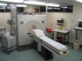
In modern PET computed tomography scanners, three-dimensional imaging is often accomplished with the aid of a computed tomography X-ray scan performed on the patient during the same session, in the same machine.